The anatomy and physiology of strokes and TIAs
You should possess a basic understanding of the anatomy and physiology (structure and function) of the blood supply to the brain. Obtaining a thorough understanding of the anatomy of the brain, as well as its blood supply, will assist you in understanding why stroke symptoms occur. Furthermore, I will be discussing about The anatomy and physiology of strokes and TIAs and you will learn how they may differ in different parts of the brain that are damaged. Learn more about the structure and function of the brain that is relevant to strokes and transient ischemic attacks by visiting the links below.
Strokes and TIAs: Anatomy and Physiology
The surface gray matter of the brain, or the cerebral cortex, makes up most of the brain. It is composed of two halves, the right and left hemispheres, which are connected by a structure called the corpus callosum. Four lobes are present on each side of your brain: the frontal lobe, the parietal lobe, the temporal lobe, and the occipital lobe.
There are different regions of the cerebral cortex in the brain that control different functions. The image below shows a visual representation of the brain’s lobes.
The frontal lobe is located in the front part of the brain and is responsible for higher mental functions, eye movement and voluntary motor function. Sensory information is processed in the parietal lobe of the brain, whereas eyesight is processed in the occipital lobe (at the back of the brain). During the processing of memories, the temporal lobe integrates sensory information such as taste, sound, sight, and touch into memories. Understanding language is also a function of the temporal lobe.
Read also ; What is stroke and TIAs

The brainstem
Between the brain and the spinal cord is the brain stem, an area at the base of the brain. As well as beating the heart, breathing, regulating blood pressure, swallowing, and other vital functions, the brain is also responsible for the body’s immune system.
The brainstem is composed of three parts:
-
Pons
-
Medulla oblongata
-
Midbrain
Review the image below, which shows the pons and midbrain. Under the pons and between the spinal cord and the medulla oblongata lies the medulla oblongata.
:max_bytes(150000):strip_icc()/human-brain-diagram-477524322-5aac1d99c67335003634c40a.jpg)
The cerebellum
There is no doubt that the cerebellum is one of the largest sections of the brain, second only to the cerebral cortex in terms of size. The brain is located in the posterior and inferior regions of the cranial cavity of the skull. As a result of its highly folded surface, the cerebellum can accommodate an increased number of nerve cells. There is an increase in surface area in the outer gray matter cortex of the cerebellum.
Cerebellum-mediated movement evaluation is the primary function of the cerebellum in evaluating the effectiveness of cerebral cortex-mediated movements. The cerebellum detects discrepancies between movements initiated by the cerebral motor area and how they are performed. The motor areas of the cortex receive a feedback signal. Allowing errors to be corrected and smooth movements, feedback signals allow complex muscle contraction sequences to be coordinated. Cerebellum play a crucial role in posture and balance besides coordinating skilled movements.
Check out the following image showing where the cerebellum is located.
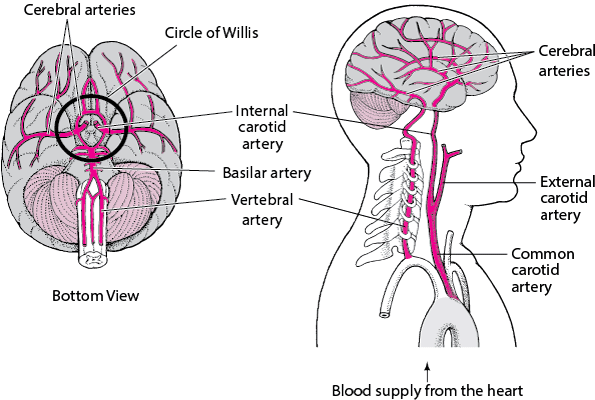
The Circle of Willis
There are two sets of blood vessels that supply blood to the brain: the internal carotid arteries and the vertebral arteries. Contrary to vertebral arteries, which run next to the spine, carotid arteries enter the brain neck through the front of the neck. Abrasions in the brain can be treated with the help of these blood vessels. The branches of these blood vessels lead to many smaller vessels.
Carotid arteries and vertebral arteries supply the brain with three major arteries:
-
Anterior cerebral artery
-
Middle cerebral artery
-
Posterior cerebral artery
At the base of the brain, these three main blood vessels form ‘The Circle of Willis’. The circle of blood vessels serves as a mechanism for compensating for the lack of blood circulation to the brain. There is a blockage in one section of the circle, which is causing this problem. The protection provided by this protection does not always fully compensate for blockages in vessels in the brain in stroke cases.
Other arteries travel from the Circle of Willis to all parts of the brain, including the anterior cerebral artery (ACA), the middle cerebral artery (MCA), and the posterior cerebral artery (PCA).
You can see a visual depiction of the Circle of Willis on the underside of the brain by watching the below video. It names each artery that arises from the internal carotid arteries and vertebral arteries.
This video is courtesy of the Royal College of Physicians and Surgeons of Glasgow.
.
If you have a stroke in various areas of the brain, what happens next?
People may experience strokes differently depending on the location of the stroke in their brains. There are several signs and symptoms characteristic of strokes that affect the blood supply of different areas of the brain in this page. These include:
-
Cerebral cortex or ‘cerebrum’
-
Right hemisphere (half) of the brain
-
Left hemisphere (half) of the brain
-
Cerebellum
-
Brainstem
Cerebrum/Cerebral cortex
-
An increase in cognitive ability (thinking, reasoning, judging, and remembering).
-
Problems with swallowing and eating
-
Problems with vision
-
Having difficulty speaking and understanding
-
Changes in sensations and movements
-
The ability to care for oneself is impaired
-
Loss or alteration of bladder and bowel control
-
Problems with emotional control
-
The functioning of sexual organs may be affected
The right hemisphere stroke
-
Problems with the visual sense
-
Visual problems and difficulties with depth perception
-
Lack of understanding of maps and inability to locate objects on them
-
The left side of the body becomes paralyzed or sensations change
-
Having trouble locating or recognizing body parts
-
Having trouble remembering things
-
Changes in behavior
The left hemisphere stroke
-
Problems with the visual sense
-
An inability to solve math problems or organize, reason, and analyze information
-
An inability to learn and apply language and information skills
-
The right side of the body is paralyzed or has sensation changes
-
Changes in behavior, like depression
-
Having memory problems
-
Unable to speak
The cerebellum
-
Having difficulty walking
-
The inability to coordinate
-
Balancing problems
-
Dizziness
-
Aching of the head
-
Vomiting and nausea problems
Brainstem
-
Feeling weak or having paralysis in the legs and arms.
-
Having difficulty swallowing, chewing, and speaking.
-
Coordination and balancing problems
-
Functions of the heart and lungs
-
There is an alteration in the control of body temperature
-
In severe cases, a coma may occur
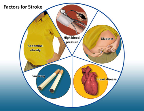
Factors that increase the risks
People are more likely to have a stroke or suffer from a TIA when they have several risk factors. Among them are:
-
People over the age of 55 are most likely to suffer strokes, although younger people can also suffer from them
-
The sex of the male
-
A previous stroke or transient ischemic attack
-
The biggest risk factor for hypertension is high blood pressure, which is preventable and treatable
-
A high level of cholesterol in the blood
-
A blood disorder called sickle cell disease
-
Taking up smoking
-
Having diabetes
-
Stroke or transient ischemic attack history in the family
-
An irregular heartbeat is called atrial fibrillation
-
Failure of the heart